24x7 Helpline Number +91-95746 25554
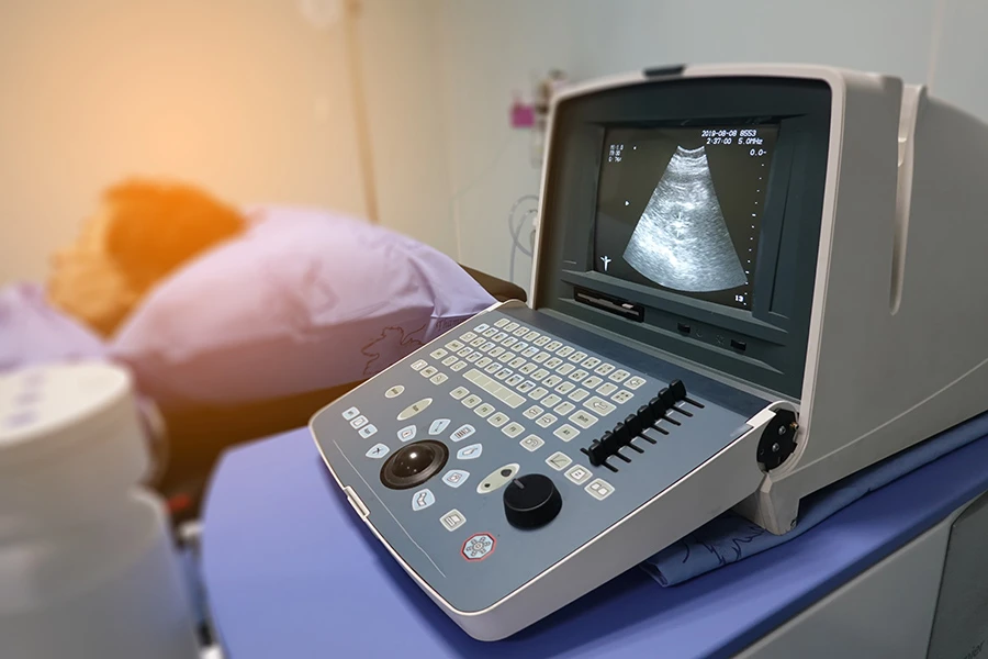
Sonography, or Ultrasound, utilizes high frequency sound waves (not x-rays) inaudible to the human ear, are transmitted through body tissues to obtain diagnostic images. Ultrasound imaging is used to evaluate many parts of the body, including the abdomen, blood vessels, superficial body structures. The echoes are recorded and transformed into photographic images.
On the day of the test:
It is very important to arrive with a full bladder. This allows the radiologist to view the bladder while it is full and after it has been emptied. Your ultrasound test is performed by registered, expert radiologists. How the Test Is Done For an abdominal ultrasound, you’ll lie down and radiologist will put a certain gel on your belly. This helps carry the sound waves. Then the radiologist will hold a probe against your belly and move it around to get an image.